Entheses
Entheses
by
Jean Luc Cornille
“Greatest scientists are always artists as well." - Albert Einstein
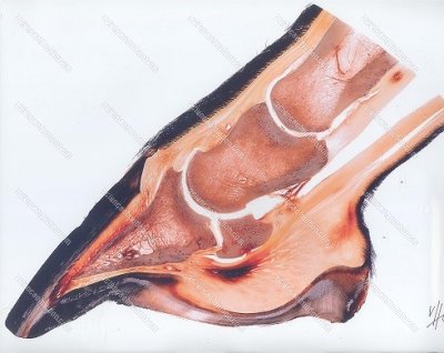
(Images by Christoph von Horst)
Dr. Christoph von Horst - www.plastinate.com
Einstein’s completed quote is, “After a certain high level of technical skill is achieved, science and art tend to coalesce in esthetics, plasticity, and form.”
The scientist who completed this “masterpiece” is undoubtedly an artist but the real beauty of the picture is that with the new generation of researches, science and art coalesce for the prevention of injuries. Dr. Betsy Uhl, DVM, PhD, is preparing the next Science of Motion International Conference, to be held October 3 and 4, 2015 at the SOM training center, and this beautiful illustration demonstrates how the study of ‘entheses,” which is the named given to the areas where tendons are inserted on bones, can prevent, the development of navicular syndrome. 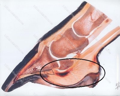
As you know, the science of motion does not believe that riders have baby brains that have to be feed with baby food. While the general consensus is repeating infantile theories, we provide instead pertinent information, often the cutting age of equine research studies. Other studies are published but the superiority and trade mark of the science of motion is the practical application. We are not afraid to question traditional beliefs in the light of new knowledge. Indeed, updating riding and training techniques to advanced scientific discoveries greatly further riders and trainers’ ability to prevent injuries.
New equine research studies focus on preventing the disease and the study that Dr. Betsy Uhl is currently developing is going to blow the mind of the ones who will attend the 2015 SOM International Conference.
Dr. Christoph von Horst - www.plastinate.com
This is the illustration of a damaged navicular bone. 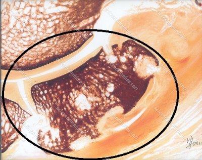 There is no need to discuss the fact that preventing such damage is thousand times more efficient than trying to deal with an affected distal sesamoid bone.
Preventing involves the capacity of interpreting early signs and the new generation of “entheses” studies provides amazing perspectives. Entheses is the name given to the area where elastic structures such as tendons, are inserted to bones. 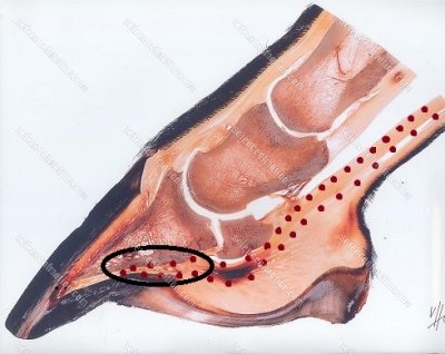 Any common sense in physics allows understanding that the connection between a solid structure such as a bone and an elastic structure such as a tendon is submitted to intense and specific forces. The attachment of the deep digital flexor tendon on the coffin bone is quite large. On the left picture we placed dotted lines around the deep digital flexor tendon. The oval shows the area where the deep digital flexor tendon inserts on the coffin bone. We place below an enlarged view of the area where the deep digital flexor tendon inserts on the bottom of the coffin bone. We surrounded the area with a discreet triangle.
(images)Dr. Christoph von Horst - www.plastinate.com
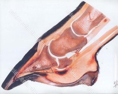 All the area below the navicular bone is loaded with receptors. There is a very large number of sensors, which is understandable since a great percentage of the forces coming from the body down to the hoof through the limb is acting on the heel area. On the next picture, the area with the very large number or receptors ia surrounded by an oval. One member of the team, who is not a rider theorized, “This is how the hoof control the gaits?” Betsy, who is an advanced rider reacted immediately placing her fingers like a hoof capsule and pushing energetically her hand down. Gravity is not acting from the hoof up into the leg. Gravity is acting downward, from the body mass down to the heel through the bony column of the limb.
The large quantity of sensors is there to measure impact forces. The dark red area that you can see just under the navicular bone is an area of the deep digital flexor tendon that is damaged by intense stress.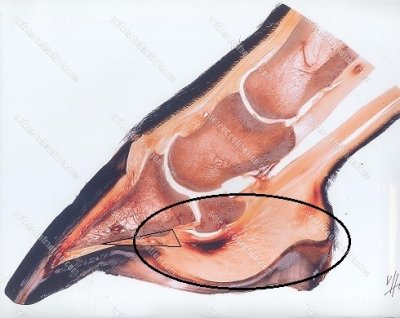 At this point, the navicular is not damaged and this is where are the value of these entheses studies. The pain is there, but x-rays will not show yet, navicular change. The horse will then likely diagnosed as having a bruised heel. It will be given butt, advised some rest, or put back to work. The warning is there. There is a kinematics abnormality of the front limb that is inducing excessive stress in the navicular area. If the kinematics abnormality is not addressed, the next step will be the development of navicular syndrome. At this point, the bone is not affected and the recovery can be total. A few months later, the kinematics abnormality will still have to be addressed but the damages on the bone will render the rehabilitation more difficult.
In his study on dancers and musicians, Boni Rietveld wrote that dancers know their body and listening to them a watching the practicing the movement and identifying the source of the kinematics abnormality allowed figuring the site of damage before advanced technologies could even show it. Horses express pain the same way but primitive riding and training techniques dismiss the information as behavior. When I saw the damage on the deep digital flexor (images)Dr. Christoph von Horst - www.plastinate.com) tendon immediately under the navicular bone, I had in front of my eyes a suspicion that was in my mind for years. I never truly believed in the diagnosis of bruised heel. I saw the discomfort lasting for a long period of time and not truly responding to drugs and hoof cares. When I was asked to deal with such problem, I always looked for the kinematics abnormality, identified the source and focused on correcting the root cause. Of course proper functioning of the hoof capsule is necessary. In fact, one of the speakers of our 2015 international conference is a very talented and sophisticated farrier who is going to explains and demonstrates how the hoof works.
For the members of the course who have watched our case study on navicular syndrome, one of the five horses, a paint horse, did not showed navicular changes on radiographies but exhibited the kinematics abnormalities that have leaded other horses to the disease. We approached the problem as if it was another case of navicular syndrome and we restored proper limb kinematics by identifying and correcting the root cause. The horse became sound and never developed later in his life any navicular issue. This horse was very probably a case where the deep digital flexor tendon was damaged as on the picture, but we corrected the kinematics abnormality before it started damaging the distal sesamoid bone.
Jean Luc Cornille 2015
To learn more about Science Of Motion International conference sign up for our free newsletter details on conference
will be published on next newsletter
Click Here to register for Newsletter Drowning The Fish


