

Hind Legs Engagement and Stifle Problem
Therapy Through Motion
Hind Legs Engagement and Stifle Problem
Equine Biomechanics
“The thought that the horse's gait could actually be changed to rehab or prevent injuries is almost completely foreign to veterinarians as well as trainers.” (Betsy Uhl, DVM, PhD. 2011)
When equine locomotion and athletic performances are analyzed in great details, like under the microscope, limb kinematics abnormalities causing injuries can actually be corrected. The horse’s physique can actually be optimally coordinated for the athletic demand of the performance. This evolution is made possible by updated understanding of the horse’s physiology.
Born with advanced research studies, the Therapy Through Motion has mature into a different field of research; the training ring. The result is a powerful therapy identifying and addressing the source of the kinematics abnormality causing the injury.
“A major cause of lameness is lameness.” Rooney’s famous idea is that when it is repeated every stride a mild kinematics abnormality causes injury. Kinematics abnormalities may originate from morphological flaw or muscle imbalance, but also from training misconceptions. Lacking the support of adequate scientific knowledge great authors’ thoughts have been distorted over time and simplified to the point of meaningless formulas. In fact simplistic formulas are the main cause of equine injuries. In this series, we review, one by one, the kinematics abnormalities causing injuries and how training misconceptions can create such abnormalities.
Hind Legs’ Engagement.
Whatever the horse’s specialty, the base of all equine athletic performance is the engagement of the hind legs. The point here is not to question the need for hind legs’ engagement but instead to underline the fact that focusing on the hoof placement is a simplification, which places the horse at risk of injury. Sound locomotion demands precise coordination between forward swing of the hind limb around the hip joint and dorso-ventral rotation of the pelvis. In his quantitative study on Swedish Warmbloods comparing back and limbs kinematics of good and bad movers, Mikael Holmström observed greater pelvis rotation on above average movers. “The undulation of the pelvis was larger in the horses with good trot and increased in passage.” (1)
The hind limbs and the pelvis have to move in the same direction. When the hind limb swings forward, the pelvis rotates dorso-ventrally. When the hind leg moves backward into the pushing phase, the pelvis returns into a more horizontal position. Pelvis and limb movements are proportional but soundness demands their precise synchronization. The problem is that it is possible through whip or spurs to create deeper engagement of the hind leg without adequate pelvis rotation. The kinematics abnormality might please uneducated eyes but places the horse at risk of sacroiliac (SI) strain and stifle problem.
There is actually a strong recurrence of SI problems. One reason might be greater concern from the veterinary world for back problems. “Even if back soreness is thought to be only a compensation for hock pain and other musculoskeletal disorders, practitioners still have an obligation to evaluate and manage the back problem concurrently.” (2) The second reason might very well be the fast forward misconception that is currently rewarded in the show ring.
Some of the kinematics abnormalities leading to sacroiliac strain are similar to the kinematics abnormalities inducing stifle problems. The kinematics of SI injuries will be studied in detail during our next Immersion Program. In this discussion, we focus essentially on the misconceptions about hind leg engagement that is placing the horse at risk of stifle injuries.
If forced to do so, a horse not using the vertebral column properly will deeply engage the hind leg underneath himself furthering the forward rotation of the femur around the hip joint. While rotating around the hip joint, the femur undergoes simultaneously an inward rotation around the tibia. This rotary movement of the femur occurs toward the outside, (medial-to-lateral,) during the swing phase and toward the inside, (lateral-to-medial,) during the support phase.
As the protracting hind leg swings forward, the stifle extends and the usual medial-to-lateral rotating movement occurs. “If the extension is carried on beyond about 143-145°, there is a final lateral-to-medial twist, which rotates the patella medially and hooks the medial patellar ligament over the medial ridge of the femoral trochlea. The stifle is “locked” and flexion prevented.”(3) This is the mechanism of accidental locking of the patella. To unlock the stifle, the quadriceps muscle contracts, lifting the patella as the biceps contracts, pulling the patella laterally. The horse’s quick reflex contractions prevent accidental locking of the patella but induce stride after stride of abnormal stresses on the joint.
At the canter, the problem is unlikely to occur because the gait does induce longitudinal flexion of the horse’s thoracolumbar spine and the pelvis does oscillate dorso-ventrally. Both hind legs are moving together into the swing phase and the axis of rotation is the lumbo-sacral junction. At the contrary, at the trot as well as at the walk, one hind limb moves forward and the other moves backward. Each limb rotates around the hip joint and the pelvis “ducktail.” The dorso-ventral rotation is naturally reduced. When training misconceptions work toward stiffening the horse’s thoracolumbar column, dorso-ventral rotations of the pelvis are reduced even more and the horse increases the rotation of the femur around the hip joint.
On a video recorded along the dressage ring of the Atlanta Olympics for kinematics studies, several horses exhibited grotesque parodies during the medium walk. The horses over tracked the hind hooves, without adequate dorso-ventral rotation of the pelvis. They were moving at the walk like Tennessee walkers. The ducktail motion of the pelvis was accentuated and they further rotated the femur around the hip joint extending the stifle joint too far. Later during the dressage test, these horses demonstrated severe stifle pain during piaff and passage. Through manipulation, one can easily create dorso-ventral rotation of the pelvis. The relation between pelvis rotation and overall flexion of the horse’s thoracolumbar spine is then apparent.
Riding techniques altering longitudinal flexion of the horse’s thoracolumbar spine, therefore expose the horse to stifle injury. The most common misconceptions that stiffen the horse’s thoracolumbar column are speed and weight on the bit. A horse increases the speed by stiffening the thoracolumbar spine. The same reflex contraction is used by a horse leaning heavily on the bit. Lowering of the neck also tends to stiffen the back. Some horses have been shown to lose vertebral mobility in the thoracic area and all horses are losing vertebral mobility in the lumbar area when the neck is lowered.
The horse’s adaptation to the rider’s weight is to increase the duration of the hind limb’s supporting phase. More exactly, the horse increases the duration of the decelerating phase. This of course demands a more forward placement of the hind leg at impact. However, efficiency does not relate to the hoof placement but instead to how well each joint of the alighting hind limb is placed to optimally absorb impact forces. At the piaff for instance, the horse places the alighting hind leg less forward under the body than during collected trot. Uneducated riders think otherwise but their opinions lack understanding of the performance’s athletic demand.
While the hind and front limbs are acting like a lever at the walk, they work more like a spring at the trot. The vertical position of the hind limb under the croup is best suited for the task of decelerating the horse’s body through the flexion of the joints. “The hind legs have a considerable braking activity to avoid forward movement of the body over the forelegs. The forelimbs have a larger propulsive activity.” (4) As the joints of the supporting hind leg fold, they resist gravity and forward displacement of the body over the forelegs. Gravity and inertia forces are loading the supporting hind leg. If the hoof was placed more forward under the body, the stress on the canon bone would be greater. Also, the hock’s middle joint T3-TC would be under excessive stress and therefore prone to arthritis. 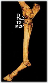
The opposite is equally damaging. When the supporting hind leg alights behind the vertical, the stress is greater on the hock’s lower joint, Mt3-T3. Basically the horse is placed in the situation of functional straight hock and is prone to injuries related to the morphological flaw. Horses are kind enough to perform even if the rider places their body into stressful positions. The price of course is lameness. More and more research studies demonstrate that cartilage issues, such as arthritis in the hock, result from abnormal stresses on tarsal bones. In most instances you see bone damage before you see the cartilage changes.
The horse adapts the hind legs’ hoof placement to the athletic demand of the performances. During the stride preceding the flying change, the horses achieving the best performances increase the length of time that both hind hooves remain on support. Basically, they increase the decelerating phase of the hind legs. “Preceding a lead change, the higher-scoring horses increased their contact duration of the hind limbs and decreased the length of step and time between forelimb impacts to prepare to execute the lead change in the succeeding airborne phase.” (5)
The race horse engages the hind legs more forward under his body than the dressage horse. Such engagement is definitively helped by the longitudinal flexion of the thoracolumbar spine and dorso-ventral rotation of the pelvis and sacrum around the lumbo-sacral junction, which is all natural at the canter. However, the race horse does not utilize greater engagement of the hind legs to enhance balance. Instead, the race horse utilizes the elastic strain energy accumulated during the decelerating phase to maximize the propulsive action.
The horse’s morphology also does influence the position of the hind hoof under the body at impact. For instance, two horses working with the same thoracolumbar column and pelvis rotation; a horse with a sickle hock will place the hind hoof more forward while a horse with a straight hock will place the hind hoof less forward. Forcing a straight-legged horse to track up deeply, would place the horse at risk of hock injury as well as sacroiliac problems and/or stifle issues.
Simplicity is the greatest achievement of knowledge but simplicity without knowledge is the greatest cause of equine injuries. “The horse’s hind legs need to track up at working trot”, is the type of simplistic formula which, if applied without sound understanding of the horse’s vertebral column mechanism and pelvis rotation, is likely to cause injury. In this circumstance the cure is knowledge. Greater engagement of the hind legs is not the cause but instead the result of sophisticated body coordination. This body coordination cannot be created by acting directly on the hind legs. Training formulas are, for a great part, grossly inaccurate and the rider’s knowledge of the underlying biomechanics factors is the horse’s best chance of soundness.
James Rooney identified the kinematics abnormalities causing injuries. The pathologist pioneered the biomechanics of lameness. The Therapy Through Motion is about identifying and correcting the source of the kinematics abnormalities causing injuries. The Therapy Through Motion pioneers the biomechanics of soundness.
Jean Luc Cornille
Copyright 2012
Our work In-Hand equine Biomechanics Course
IHTC in-hand therapy dressage course
References,
(1) (Mikael Holstrom, Quantitative study on conformation and trotting gaits in the Swedish Warmblood riding horse. Dissertation, Uppsala, 1994)
(2) (Kevin K. Haussler, DVM, DC, PhD, Preface, Veterinary Clinics of North America 1999)
(3) (James R. Rooney, Biomechanics of lameness in horses, 1976)
(4) (Erid Barrey, Sophie Biau, Locomotion of dressage horses, Conference on Equine Sport Medecine and Science, 2002)
(5) (N. R. Deuel, PhD: J. Park, PhD, Canter lead change kinematics of superior Olympic dressage horses, 1990)
Science Of Motion
Jean Luc Cornille
scienceofmotion.com
