

Stretching and Relaxation
Stretching and Relaxation
Myths and Reality
Jean Luc Cornille
BUY PDF FILE OF THIS ARTICLE TO PRINT OUT $9.95
After contraction comes relaxation. Both aspects of muscular work create movements. The quality of the movement is not proportional to the relaxation, but the intensity and timing of both the contraction and the relaxation is. In most instances, the relaxation is not a muscular slackness, but instead, a return to the initial muscle tones. This is part of the stabilization mechanism which rules proper functioning of the equine vertebral column. The same mechanism stabilizes the human’s vertebral column. “The reflex contractions of the spinal column muscles compensate for the bending of the spinal column. This is a characteristic behavior of the spine stabilizing system during human movement.” (Obber, J. K. 2002)
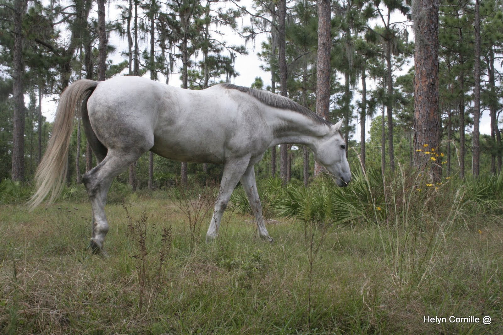
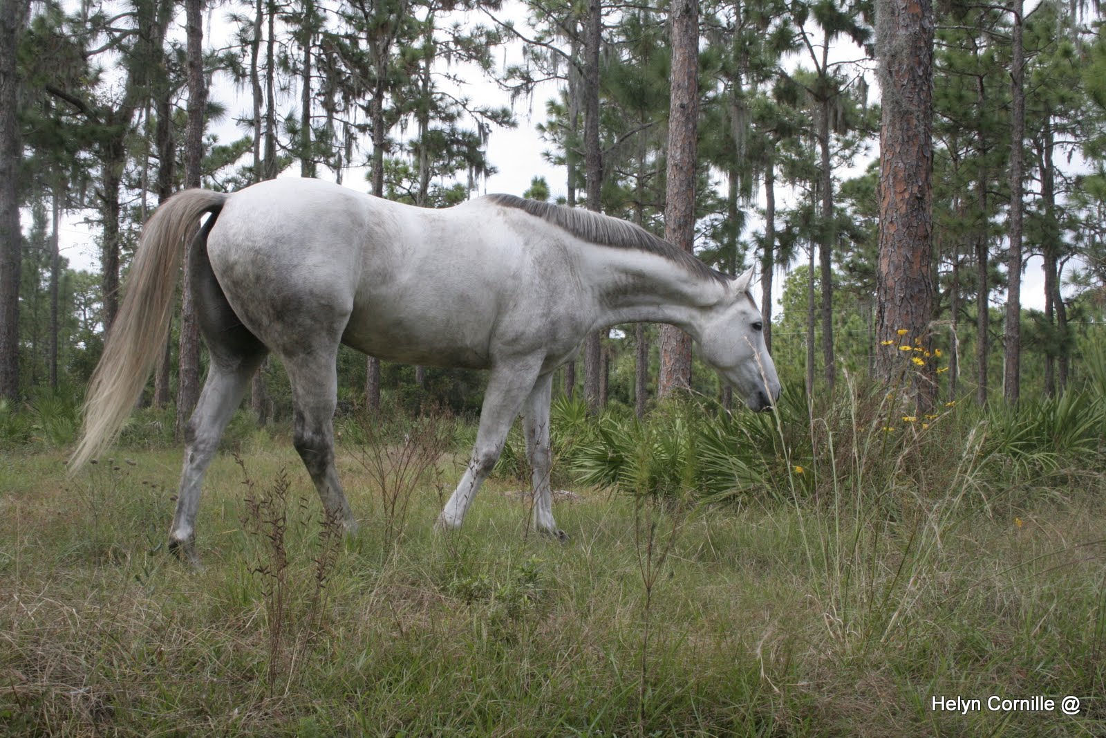
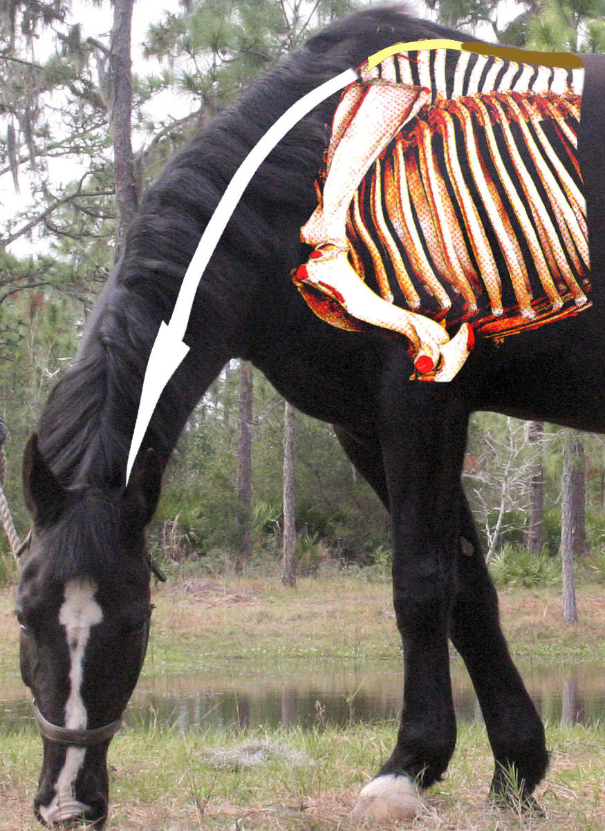
After a concentric contraction, the type that involves a shortening of the contracting muscles, the muscles then relax and return to their initial length. Technically, the muscles “stretch” as they elongate. In the case of lowering of the neck, the upper neck muscles elongate beyond their initial length. This is where myth is separated from reality.
A lowering of the neck is in fact a flexion, which is achieved by a concentric contraction of the lower neck muscles, and is aided in the task by the force of gravity that pulls the head and neck down toward earth. The upper neck muscles, situated above the cervical vertebrae, elongate to allow the lowering of the neck, but also contract to support the burden of the head and neck. When a horse is under heavy sedation, the upper neck muscles do not resist gravity and the horse’s head hangs a few inches above the ground at the limit of the nuchal ligament’s elastic compliance.
The work of muscles that elongate while contracting is referred to as “active stretching”. The work of the upper neck muscles is also called “eccentric contraction”. In terms of muscular work, eccentric contraction is the most powerful type of muscular contraction. “Muscles working eccentrically can absorb up to 15 times more energy than during concentric contractions.” (R. C. Payne, P. Veenman and A. M. Wilson, 2004)
The myths suggest that as the horse lowers his neck, the upper neck muscles stretch while relaxing. But in reality, the upper neck muscles elongate as well as support the burden of the head and neck, which is as much as 10% of the horse’s body weight. Hence, they do not stretch and relax, but work in active stretching, i.e., eccentric contraction.
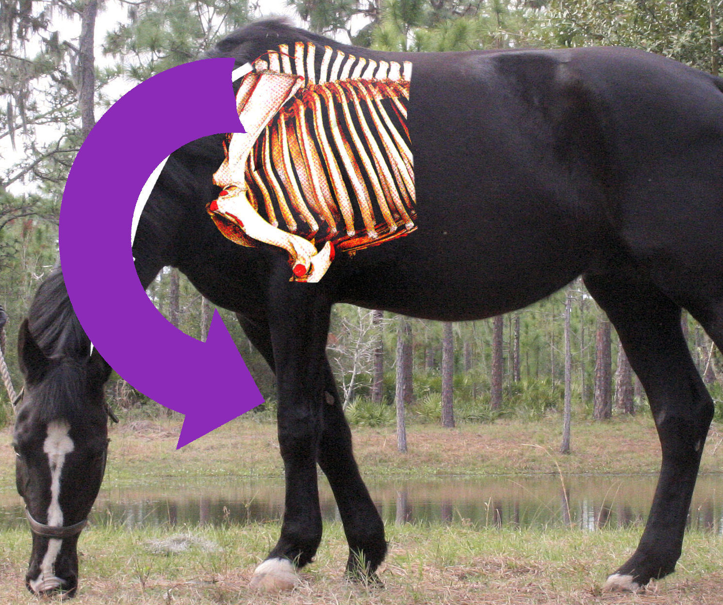
“Everything should be made as simple as possible but not simpler.” (Albert Einstein) Stretching theories are simpler. They involve two main muscle groups, the Longissimus dorsi, which connect the sacral vertebrae to the middle of the neck, (cervical vertebrae,) and the Spinaleus dorsi which extend from the wither back along the vertebral column. Both terms are generic. The longissiumus system is in fact composed of several muscles, longissimus cervicis, capitis, lumborum, ect. Oriented in mirror image direction is the spinaleus system that refers to muscles such as the multifidius which fascicles are oriented downward and backward. Downward, means that the fascicles are oriented from the dorsal spinous processes down to the body of a vertebra situated three to five vertebrae behind. Backward, means that the fascicles are oriented toward the back.
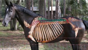
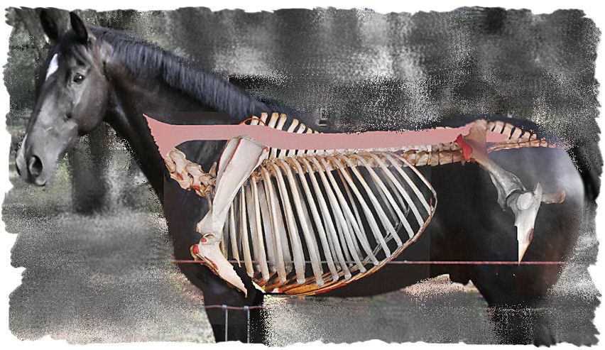
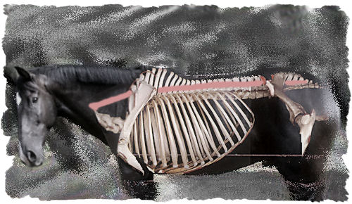
Simplistic representations illustrate the longissimus system as a long and thick bungee cord stretching from the sacrum to the fourth cervical vertebra. Based on such schematic illustration simplistic thoughts deducted that the lowering of the neck elongated the horse’s whole top line.
A slightly more elaborated illustration of the longissimus system as the one presented in 1946 by the Dutch scientist L.J. Slijper, demonstrates that such simplistic theory does not apply to the back muscles’ structure. The longissimus dorsi muscle group is composed of fascicles bridging three to five vertebrae.
Therefore even if the upper neck muscles were pulling down and forward on the back muscles, as claimed by stretching theories, the effect would not go further back than the whither area.
Furthermore, if the upper neck muscles were pulling on the back muscles down and forward, they would not be able to support the burden of the head and neck. The horse’s head would then hang a few inches above the ground at the limit of the nuchal ligament’s elastic compliance, as demonstrated by the horse under heavy sedation.
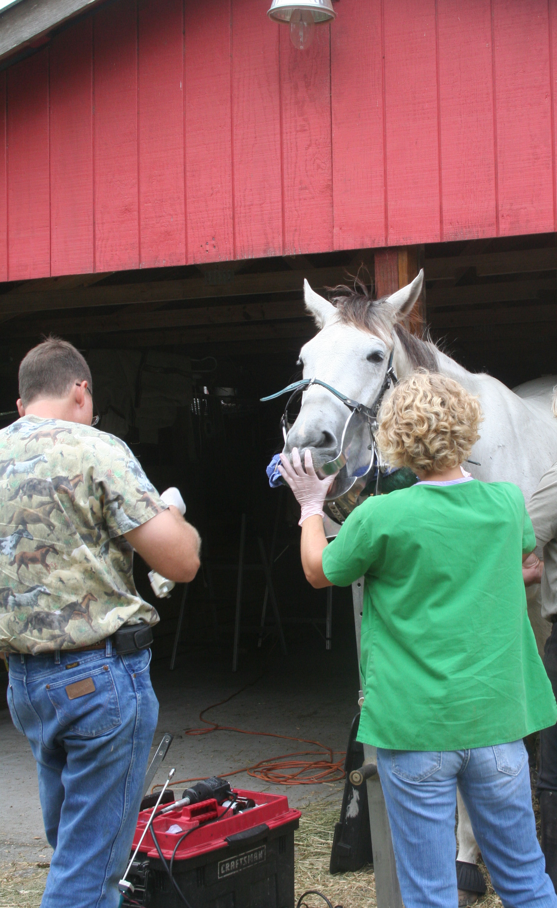 On the picture, the horse is prepared for the dentist and the effect of the sedation is only starting.
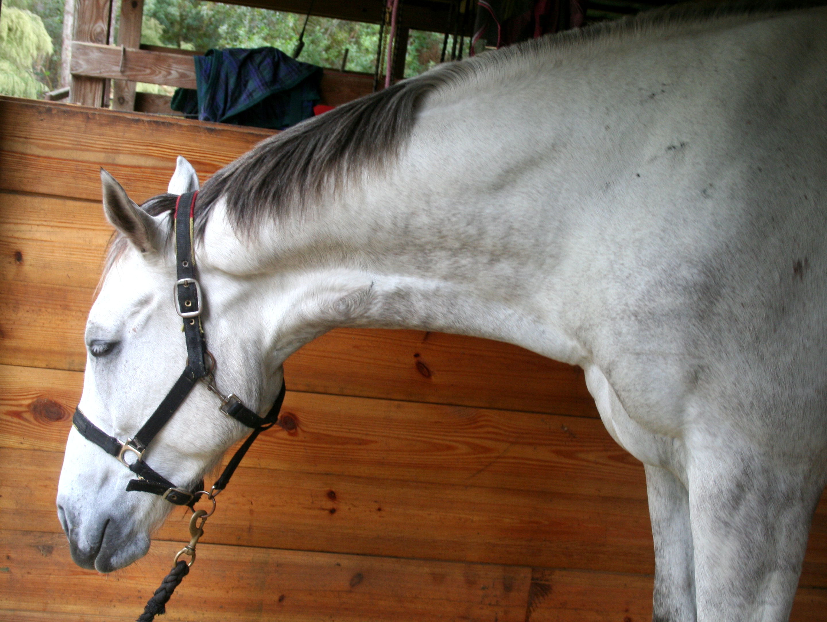
The same horse holding a similar neck posture while not under sedation shows a very different work of the upper neck muscles. 
Even myths have their logic, however, and the belief that the lowering of the head and cervical vertebrae pulls on the back muscles, thereby stretching and relaxing the horse’s top-line, is supported by the feeling of ease that is often associated with work with a longer neck posture. The horse’s vertebral column does feel more “round” and at ease when the neck is held in a longer posture. The problem is not the accuracy of the rider’s perception but rather the translation of the perception. The feeling of ease does not result from relaxed elongation of vertebral column muscles but instead from the way the vertebrae rotate in relation to each other. The phenomenon is known as “Instant Center of Rotation.”
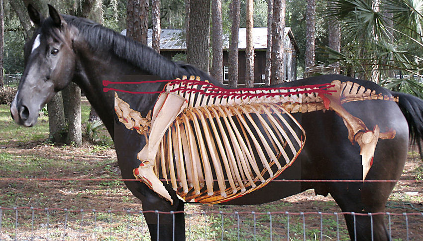
Vertebrae rotate around each other, and if a study is made of just two vertebrae of the equine spine, one would focus on an area that scientific research defines as inter-vertebral joint complex, or motion segment. 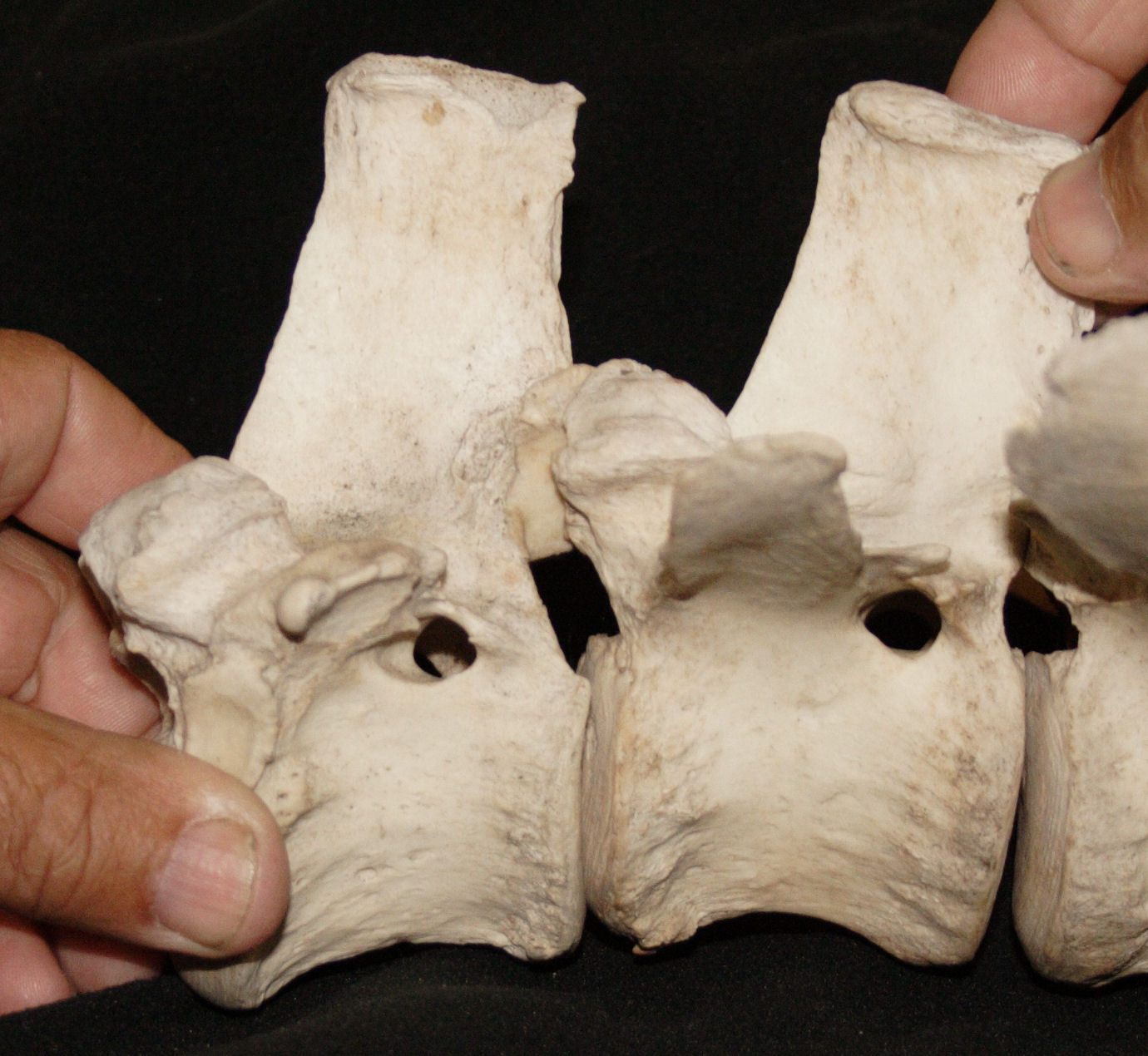
“The functional unit in spinal kinematics is the ‘inter-vertebral joint complex’ or ‘motion segment’, which is composed of two contiguous vertebrae and their associated soft tissues.” (Denoix)
Using a geometric model, the rotation arc of one vertebra around the other can be described as the segment of a circle. The axis of rotation of the rotating vertebra is situated toward the middle of the previous vertebral body. 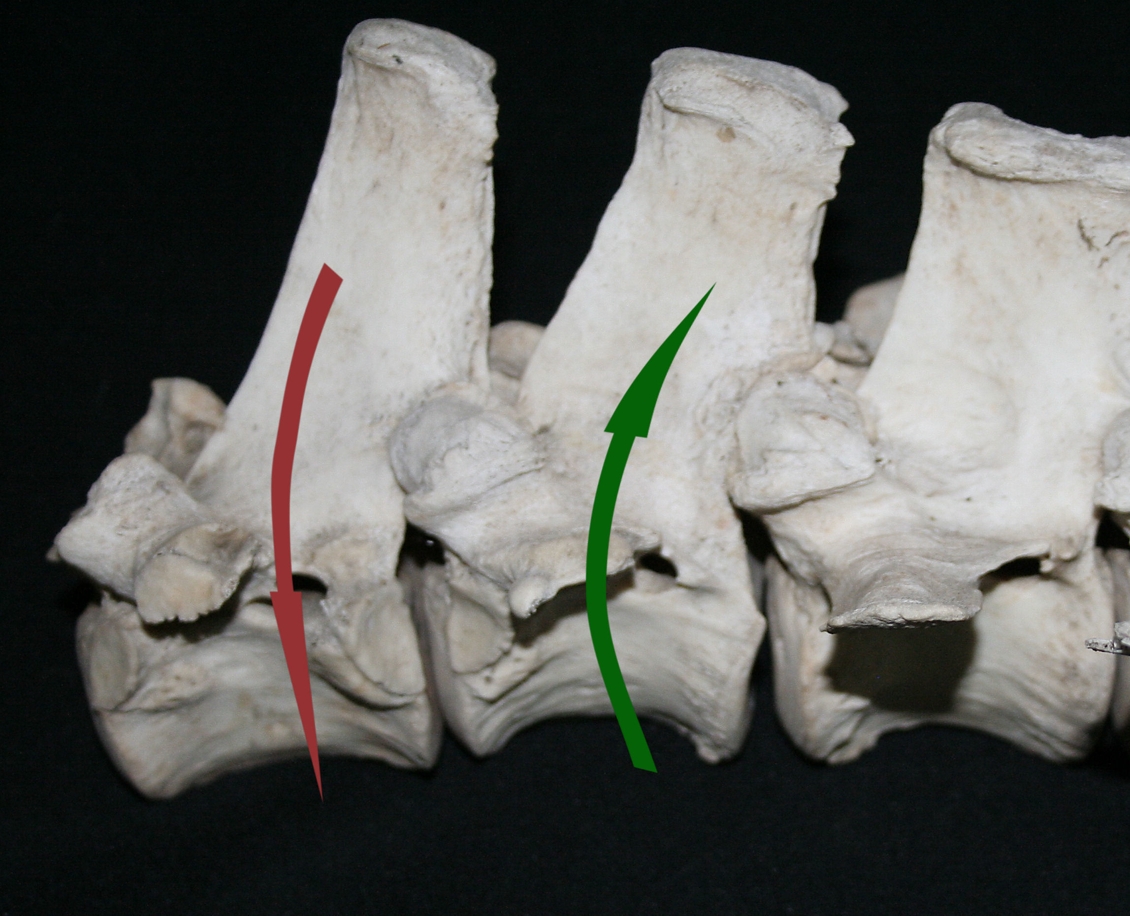
On this picture, the vertebral rotation that creates longitudinal flexion of the spine is illustrated in red. The vertebral rotation that creates extension of the spine is illustrated in green.
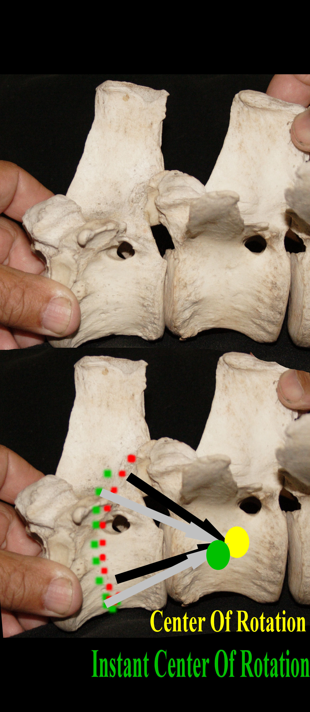
The theoretical center, wherein one vertebra rotates around the other can be labeled “center of rotation”. 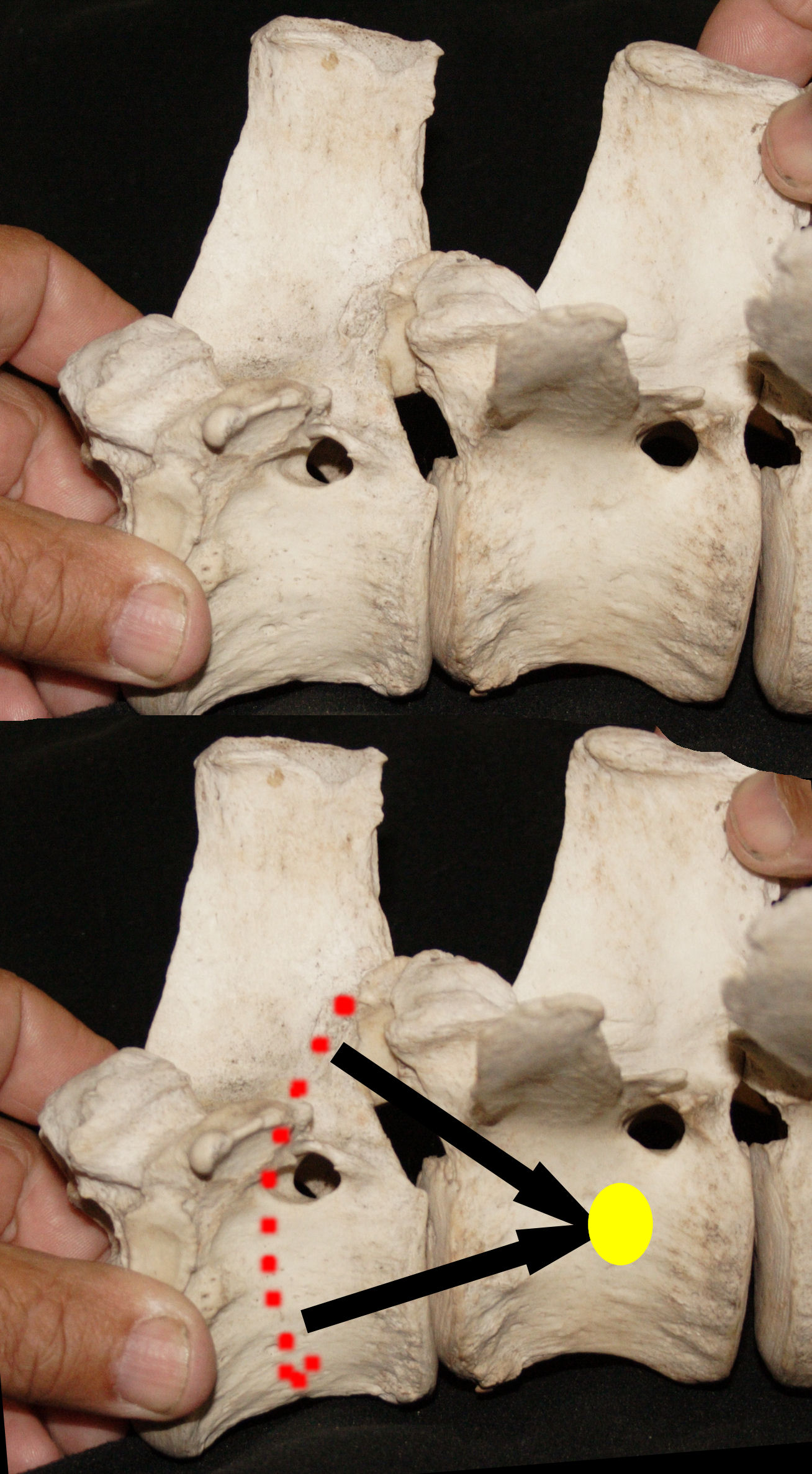
In our example, however, the central vertebra is fixed, while in motion, this vertebra is also rotating around the previous vertebra and therefore, the center of rotation can only last an instant. This is why it is labeled “instant center of rotation”.
When the neck is lowered, the instant center of rotation moves down and forward toward the inter-vertebral disk.
If one compares the rotation of one vertebra around the other as the horse’s neck is held in a natural posture,
with the rotation of the same vertebrae when the neck is lowered, we can observe less pressure on the lower end of the inter-vertebral disk. “The inter-vertebral disk undergoes less tangential displacement (shearing) than during thora-columbar flexion alone”.(Denoix)
The pressure on the inter-vertebral disk is quite impressive. This picture illustrates the bulging that occurs in the lower end of inter-vertebral disc during flexion of the lumbo-sacral junction. 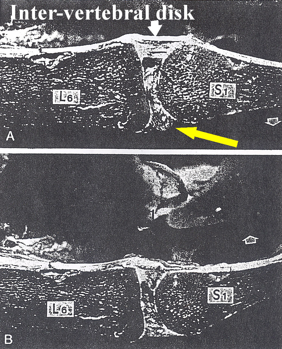
The yellow arrow is directed toward the bulging of the intervertebral disk .
The vertebral body on the left is the 6th Lumbar vertebrae and the vertebral body on the right is the first sacral. For comparison, the same vertebral segment is pictured during extension of the spine. (Lower part of the picture.) The intervertebral disk is then stressed differently
Watching the stress that a simple flexion of the vertebral column induces on the lower end of the inter-vertebral disks may help to understand the salient function of the back muscles. The whole muscular system surrounding the horse’s vertebral column is designed to primarily protect the spine from intensity and amplitude of movements that would exceed the spine’s integrity and possible range of motion.
Greater stress on the vertebral structure stimulates stronger protective reflexive contraction of the surrounding muscles, while less strain incurs less muscular intensity. The feeling of ease associated with a lower neck posture may results from the adjustment of the epaxial spinal musculature to lesser stress on the vertebral structure. Lesser contraction might be described as relaxation but the meaning is quite different from the concept of relaxation that suggests looseness and elongation of the back muscles.
Nicole Barthel, who translated General Decarpentry’s Academic Equitation into English wrote in her preface, “Relaxation: The French use the word ‘decontraction’, as the opposite of contraction. From the point of view that concerns us in equitation, I would have preferred this term to ‘relaxation’, but it would not have been condoned by English linguists. Wherever the word ‘relaxation’ occurs, it must be understood that it signifies an absence of sustained contraction and not a total slackness of muscles”. (The French definition of “decontraction” is absence of unnecessary contraction)
There have been many instances in history where it was demonstrated subsequently that a situation was not as perceived. The feeling of ease associated with lowering of the neck does not result from looseness and elongation of the back muscles. Instead, the feeling of ease results from better orchestration of numerous and minuscule muscle contractions and compensatory contractions that create the rotations of the vertebrae. As well, gaits and performances cannot be enhanced increasing the horse’s vertebral column’s range of motion. Rather, performances and soundness rely on the rider’s ability to properly coordinate the minute motions of the horse vertebral column.
The perception of a rounder back is the outcome of the thoracic flexion that can be created by a slight lowering of the neck. “The lowering of the neck provoked flexion all along the thoracic spine.” (Jean Marie Denoix, DVM. PhD, 1999) However, the thoracic flexion does occur only if the lowering of the neck is treated as a cervico-thoracic flexion, which is the combined work of the muscles suspending the trunk from the front legs (Thoracic) and a longer and consequently slightly lower neck posture (Cervical).
The difference between cervico- thoracic flexion and lowering of the neck is illustrated here. Within approximately the same neck posture, the horse sustains the trunk between the shoulder blades, (left picture.) The horse sags then the trunk between the forelegs, (right picture.)
We superimposed the two pictures placing on the back the horse sustaining the trunk higher between the forelegs.
The yellow doted line follows the top line of the horse sustaining the trunk higher between the forelegs. 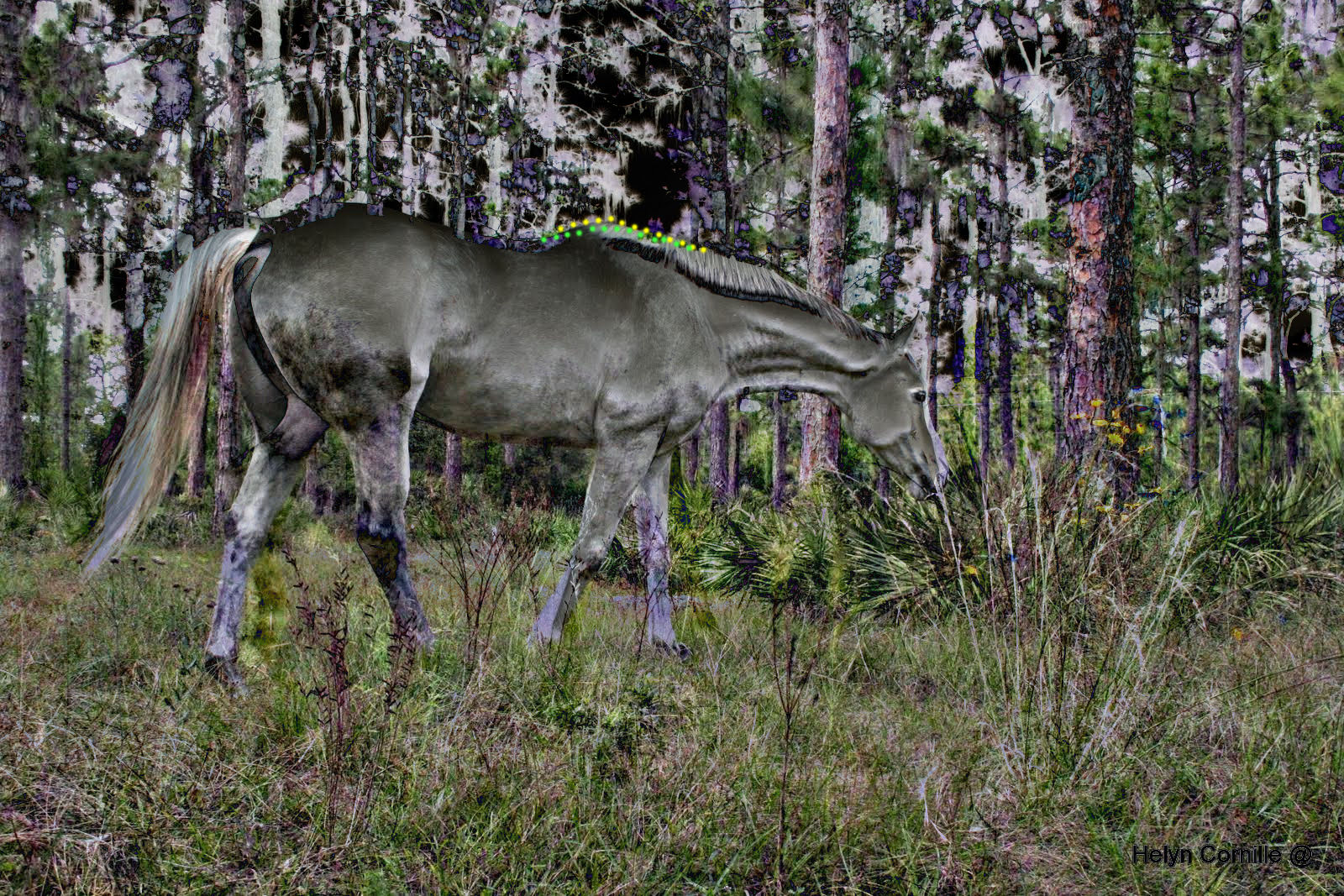
The green doted line follows the top line of the horse sagging the trunk between the front limbs.
Due to the traction that the nuchal ligament exerts on the tip of the dorsal spinous processes of the wither’s vertebrae, the lowering of the neck induces a “verticalisation of the dorsal spinous process”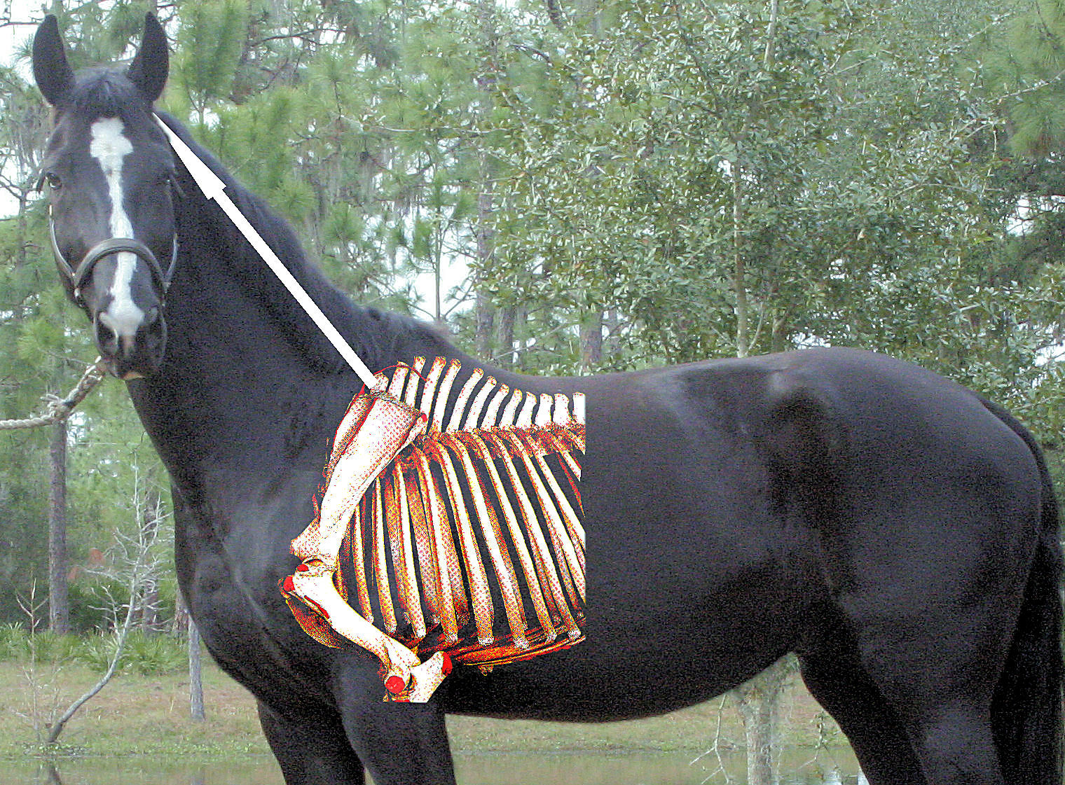
If the horse sustains the trunk between the forelegs, (Cervico-thoracic flexion), the verticalization of the dorsal spinous processes of the wither induces flexion of the thoracic spine.
By contrast, if the horse lowers the neck without adequate work of the muscles supporting the trunk from the front legs, the lowering of the neck cumulates damaging effects, increased weight on the forelegs, lost of mobility of the lumbar vertebrae, restricted dorso-ventral rotation of the pelvis.
The father of modern equitation wrote: “Theories teach us to base our work on sound principles, and these principles, rather than being opposed to what is natural, must serve to perfect nature with the aid of art”. (Francois Robichon de la Gueriniere, 1731) Riding and theirs professing greater amplitude of the horse’s vertebral column movements via stretching and relaxation are opposed to what is natural. Perfecting nature demands a subtle orchestration of the myriad minute muscle contractions and compensatory contractions that are the source of all body movements. “The biomechanics of the vertebral column, although very complex, are of vital importance because they form the basis of all body’s movements,” (Leo B. Jeffcott, 1980)
The subtle orchestration of the minute and numerous muscle contractions and compensatory contractions creating the work of the horse’s vertebral column is mostly created by the horse’s central nervous system, the brain. It would be impossible to stimulate and coordinate such complex mechanism through the traditional concept of “stimulus response”. Guiding the horse’s brain toward the most efficient body coordination demands understanding and respect for reality. The horse’s brain may eventually explore muscular coordination beyond the scope of natural reflexes, but will resist amplitude of movements which exceed his vertebral column’s possible range of motion. Hence, the horse’s brain will protect his spine from stretching, muscles’ elongation, and relaxation in the sense of loosing muscle tone.
The amplitude of the vertebral column’s movements is naturally very limited and the main function of the back muscles is to maintain the movements of the vertebral column within the limits of the vertebral column’s range of motion. “Electromyographic studies and movements data presented above strongly suggest that the primary function of the back muscles during walking is to control the stiffening of the back rather that to create movement.” (Hans Carlson, 1979)
These are the realities that the horse’s brain is designed to protect and efficiency demands that riding and training principles adjust to reality. In motion, considerable forces are induced on the horse’s vertebral column. There are the forces created by the legs, accelerations of gravity, inertia, rider’s movements, etc. The vertebral linkage is designed to absorb, convert, and redirect these forces while simultaneously maintaining amplitude and intensity of the vertebral column movements within the limits of the vertebral column’s range of motion.
Fundamentally, a large amount of forces have to be absorbed and minimized within the limits of the equine vertebral column’s range of motion. Simultaneously, great deals of diverse and minute movements have to be orchestrated within these limits. “The amount of joint range of motion at any vertebral motion segment is small, but the cumulative vertebral movements can be considerable.” (Kevin K. Haussler, DVM, DC, PhD, 1999) Therefore the work of the back muscles is to reduce forces and movements through supple resistance.
Our ancestors believed that the movements that they perceived on the saddle were the motions of the horse’s vertebral column. Until technology demonstrated otherwise, such belief was effectively the most rational explanation. Today’s measurements demonstrate that a large percentage of the movements, or “ kinematics”, that the rider perceives on the saddle are in fact perceptions of forces, or “dynamics”. “Kinematics is the geometry of movement, a graphical record of the initial, intermediate, and final positions of the various parts and pieces of the system one in relation to the other. Dynamics, on the other hand, is concerned with the forces applied to and generated by the system and, therefore, includes acceleration, mass, potential and kinetic energy, momentum, etc.” (James R. Rooney,DVM, 1969)
 This diagram shows the amount of vertical forces impacted on the rider’s physique at a slow trot. The dots moving the most are situated under the rider’ seat. Each dot represents a sensor, (accelerometer), that was fixed on the horse’s back. The line connecting the dots illustrates the forces placed on the horse’s thoracolumnbar spine during locomotion. The computer program was set to measure exclusively the vertical forces.
 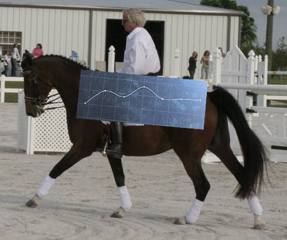 The area that is subjected to the largest amount of vertical force is the area where the rider is seated. The sensors record the sum of the vertical forces, which includes the upward propulsive forces developed by the front and rear legs, as well as the vertical forces created by the rotation of the vertebrae.
Under normal circumstances, the forelegs develop 57% of the vertical forces while the hind legs produce only 43%. These measurements focus exclusively on the limbs’ action. “In horses, and most other mammalian quadrupeds, 57% of the vertical impulse is applied through the thoracic limbs, and only 43% through the hind limbs.” (H. W. Merkens, H. C. Schamhardt,G. J. van Osch, A. J. van den Bogert, 1993).
To date, measurements differentiating the action of the limbs and the vertical forces created by the rotations of the vertebrae have not been achieved. We have observed through our approach that it was effectively possible to modify the horse’ limbs kinematics through specific orchestration of the horse’s vertebral column mechanism. However, such control demands adapting riding principles to actual knowledge. Principles of riding such as driving the horse onto the bit and swinging motion of the rider’s lumbar vertebrae, (doughy seat), are based on elementary knowledge of the equine physiology. These principles do not permit any sophisticated control of the horse’s vertebral column properties.
By contrast, riding principles based on actual knowledge of the equine physiology promote reducing the sum of the forces induced onto the rider’s physique by the horse’s movements through supple resistance of the rider’s vertebral column muscles. “The subtle S-curve of the spine allows the spine to oscillate minutely, a movement so tiny hat it is hardly perceptible to the naked eye, producing a “soft” seat. This “soft seat” differs fundamentally from a “doughy” seat, in which we find a spine that is too flexible and allowed to undulate freely in response to the horse’s movement.” (Waldemar Seunig).
Already in 1964, equine research studies suggested that the rotations of the vertebrae were converting the thrust generated by the hind legs into horizontal forces, (locomotion), and vertical forces, (resistance to gravity), “An initial thrust on the column is translated into a series of predominantly vertical and horizontal forces which diminish progressively as they pass from one vertebrae to the next” (Ricahrd Tucker, 1964). The findings strongly suggest that equestrian theories such as the lowering of the neck were too primitive. Even if the lowering of the neck had an effect on the back muscles, it would be a general action. Instead, efficiency in forward movement and balance control demands the faculty to discriminate specific areas and movements. The rider’s back, rather than any neck posture, influences efficiently the horse’s vertebral column mechanism.
In 1946 E. J. Slijper investigated the insertion of the main back muscles on the dorsal spinous processes of the vertebrae. The Dutch scientist may not have been the first to think that the muscles were rotating the vertebrae through their action on the dorsal spinous processes, but he was the first to thoroughly assess the hypothesis. Slijper compared many different species in order to soundly determine the function of each muscle. The scientist even raised a goat which was born with only two hind legs. Later, Slijper euthanized the two legged goat and compared the angle of the goat’s dorsal spinous processes by comparison with the dorsal spinous processes of a normal goat. The bipedal goat allowed him to demonstrate that his hypothesis was right. Slijper believed that the angle of the dorsal spinous processes of the vertebrae was influenced by the action of the muscles acting on the dorsal spinous processes.
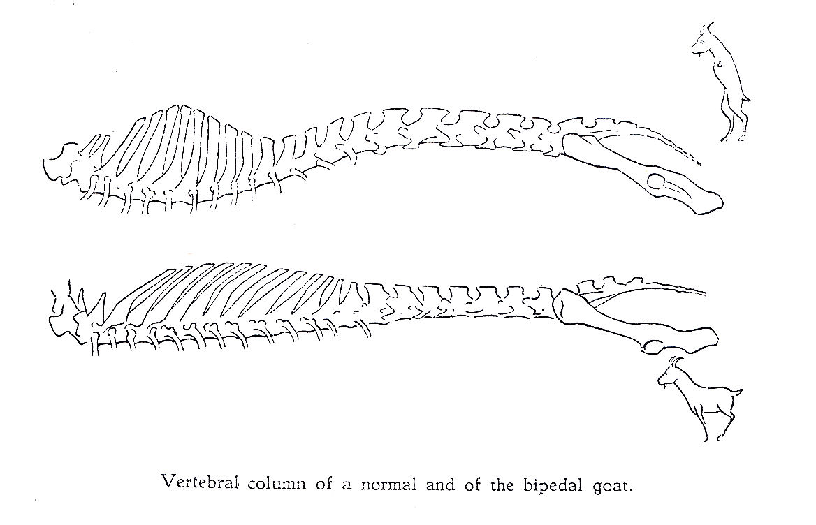
Slijper’s diagram compares the vertebral column of the bipedal goat with the vertebral column of a normal goat.
Slijper’s work demonstrated similarities between all terrestrial mammals but also offered specializations related to the animal’s size, gaits, and survival needs. Slijper (1946) already established, as did James Rooney (1969), Kevin Haussler (1999) and others, that the main back muscles were set and functioning in opposite direction.
Therefore, vertebral rotations are created, measured, compensated, and specialized, by the subtle coordination of muscles acting in opposite directions. 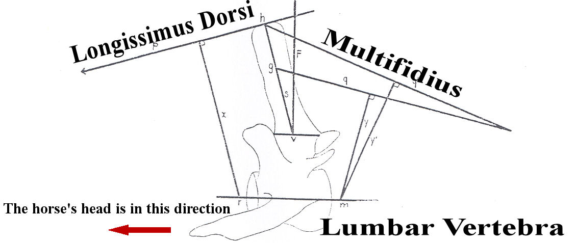
The findings should have raised a few rational thoughts. First, it is very unlikely that a neck posture acting on the back muscles in a single direction would be able to coordinate muscles groups working in opposite directions. In the same line of thought, riding principles emphasizing shifts of the rider’s weight acting back to front are unlikely to properly coordinate muscles groups such as the main muscles of the horse’s back, which are acting in opposite directions.
The topic of this series, “a failure of Olympics dimension,” offers that by failing to evolve with recently advanced knowledge of equine physiology, riding and training techniques are leaving equine athletes unprepared for the performances.
Riders’ skills and horses’ talents deserve better riding and training techniques than driving the horses into an overly flexed neck. Many great riders have found efficient solutions but their legacy cannot be understood as long as one’s thinking remains at the level of driving the horses onto the bit.
The purpose of this series is to provide the knowledge to discriminate riding and training principles that efficiently prepare the horse’s physique for the performance from riding principles that fail the horse’s talent and exploit the horse’s generosity until there is impairment.
Jean Luc Cornille
References;
- ( Obber J. K. 1074, A dynamic concept for the diagnosis of idiopathic scoliosis. In: Biomechanics IV. Proceedings of the 4th International Seminar of Biomechanics. Eds R. C. Nelson & C. A. Moorhouse, Mcmillian Press Ltd, London & bastingstoke)
- (R. C. Payne, P. Veenman and A. M. Wilson, The role of the extrinsic thoracic limb muscles in equine locomotion, 2004. J. Anat. 205 pp479-490)
- (Jean Marie Denoix,DVM, PhD, Spinal Biomechanics and Functional Anatomy, The Veterinary Clinics of North America, Back problems, Volume 15. Number 1. April 1999)
- (Nicole Barthel, Translation of General Decarpentry’s Academic Equitation, 1971)
- (Francois Robichon de la Gueriniere, Ecole de Cavalerie, 1731)
- (Leo B. Jeffcott, Natural rigidity of the horse’s backbone, 1980)
- (Hans Carlson, Halberstma, J. and Zomlefer, M. 1979, Control of the trunk during walking in the cat, Acta physical, scand. 105,251-253)
- (Kevin K. Haussler, DVM, DC, PhD, Anatomy of the Thoracolummbar Vertebral Region, 1999)
- (James R. Rooney, Biomehcanics of lameness in horses 1969,The William & Wilkins Company, Baltimore)
- (Richard Tucker, Contribution to the Biomechanics of the vertebral Column, Acta Thoeriologica, VOL. IX, 13: 171-192, BIALOWIEZA, 30. XL. 1964).
- (E. J. Slijper, 1946. Comparative Biologic-Anatomical Investigations on the Vertebral Column and Spinal Musculature of Mammals. Institute of veterinary anatomy of the state university, Utrecht, Holland. Tweede Sectie, Deel XLIL, Nº 5)
- (Xenophon, 430- 355- Before JC)
- (Leonardo da Vinci, 1452-1519)
- (Francois Robichon de la Gueriniere, Ecole de Cavalerie, 1733)
- (General Decarpentry, L’Equitation Academique, 1949)
- (Mikael Holmström, Quantitative studies on conformation and trotting gaits in the Swedish Warmblood riding horse, Dissertation, Uppsala, 1994)
