Technical discussion with Leanne
Technical discussion with Leanne
(Working Student at our farm)
The subject of yesterday technical discussion was “lowering of the neck.” As usual we approach the issue from the perspective of the horse’s functional anatomy. I placed the main upper neck muscles on the vertebral column that is suspended in my office explaining how they work. If you remember, this was the subject of IHTC3 and 4. Leanne asked me about the other muscles involved in the lifting and lowering of the neck. I told her those are the main upper neck muscles involved in lifting and lowering the neck, the Splenius, that is illustrated on this diagram with oblique lies and the Semispinalis Capitis that is illustrated in this diagram as a grey area.
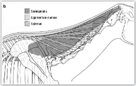
Diagrams by Michael A. Simmons.
I realized then that Leanne was referring to these illustrations that are often published showing muscles inserted in oblique for the thoracic dorsal spines to the cervical vertebrae that appears to elongate when the neck is lowered. I show her this picture asking, “Those are the muscles that you are talking about?” and Leanne acquiesced. Leanne does not believe in lowering of the neck but her objective mind directed her question. These illustrations are regularly promoted and she needed to ask about it.
On 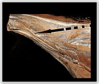 the picture, the black arrow does not illustrate the direction of muscles action. The black arrow illustrates the “stretch” that the stretching theories want you to believe. These drawings are not showing the horse’s anatomy. Instead, they manipulate the horse anatomy to fit their stretching theories. The drawings illustrate more or less the lamellar elements of the nuchal ligament. You can see the lamellar elements of the nuchal ligament on the lower left picture. Fake diagrams also illustrate the cervical elements of the serratus muscles that you can see here on the right picture.
You draw a compost of both and you can have a picture showing fascicles that would stretch if the neck was lowered. The problem is that they are not the muscles that lower of lift the neck. The nuchal ligament comes effectively under tension and elongate when the neck is lowered but this is not a muscle. This is a ligament. It assists the work of the upper neck muscles but does not create lifting or lowering. The role of the nuchal ligament is easing the work of the upper neck muscles. Gravity is pulling the head and neck down and the upper neck muscles resist attraction of gravity. Their work is helped by the nuchal ligament that is reducing the work of the upper neck muscles by up to 55% at the walk and between 32 and 36% at the trot and canter. The nuchal ligament does not fully replace the work of the upper neck muscles. The ligament just ease the work of the upper neck muscles, the muscles still work resisting attraction of gravity.
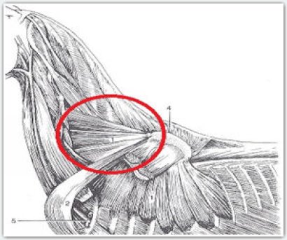
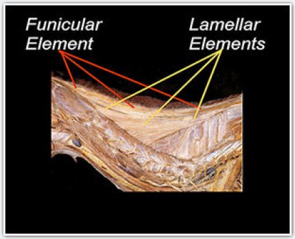
The cervical elements of the serratus muscles support the trunk between the forelegs. The muscles are also involved in raising the thorax, and the retraction of the foreleg through rotation of the scapula. They are not directly involved in lifting or lowering the neck. They are indirectly involved in the sense that the lowering of the neck increases the load on the forelegs and therefore the cervical elements of the serratus contract harder resisting greater load. Even if there is some elongation, this is not a stretching; it is an eccentric contraction, resisting elongation. Eccentric contraction is the most powerful type of muscle contraction.
The direction of the fascicles that is suggested by diagrams promoting lowering of the neck is not even close from the direction of the fascicles composing the muscles that are effectively resisting lowering of the neck. This picture illustrates the direction of the muscles fibers theorized by proponents of the stretching theories, and on these diagrams are the direction of the mu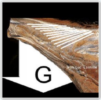 scles that are effectively controlling lowering of the neck.
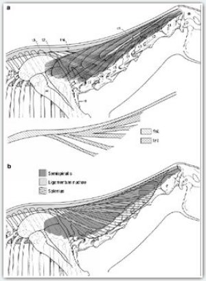
The discussion continued then on the positive effect of the lowering of the neck. There is effectively a position where the cantilever effect of the head and neck does enhance longitudinal flexion of the thoracic vertebrae. This has nothing to do with stretching or "meditation," but instead with physics. However the positive neck position is long but not low and is different for each horse. The second part of our technical discussion will be published tomorrow or the following days. You will then understand the purpose of the introductory picture.
Diagrams by Michael A. Simmons. Jean Luc Cornille 2013
PART TWO
Technical discussion with Leanne
Part 2
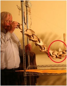
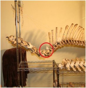
The horse’s head and neck weight approximately 10% of the horse body mass.
My right arm can be considered as illustrating the horse’s head. My hand is the poll. The weight of the head is pulled down by attraction of gravity converting the cervical vertebrae into a “loaded beam.” The shape of the beam, which is composed of the cervical vertebrae is always a S shape. The curves of the S shape might be more accentuated when the neck is held into a higher position, or closer from a straight line when the head and is placed on a lower position. Even when the horse is grazing, the cervical vertebrae can reach an almost horizontal alignment but do not revert their S shape. The red circle surrounds the cervico-thoracic junction. 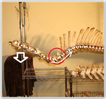
The difference between the cervical alignment of the left picture and the lower cervical alignment of the right picture results from the lowering of the neck. The head is pulled down by gravity lowering the neck. The alignment created by the S shape of the cervical vertebrae and the curvature of the thoracic vertebrae is enlightened on this picture by the white dotes. Due to this alignment, the lowering of the neck created by the weight of the head, does have a lifting effect on the thoracic vertebrae . However, this is not a systematic effect. The lowering of the neck does induce thoracic flexion, but only into a specific position that is proper for each horse and under precise conditions.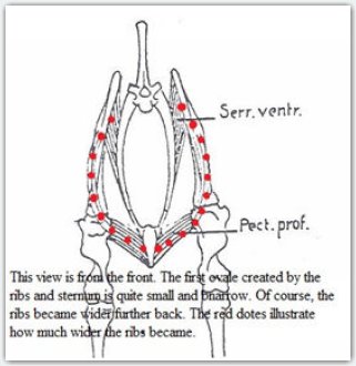
The trunk is suspended between the forelegs by a network of muscles. The most important is the serratus ventralis thoracic and cervicis. The illustration presented here is from L. J. Slijper in 1946. At this time Slijper believed that the pectoralus profondus was major in supporting the trunk. Greater understanding of functional anatomy, as well as muscles’ architecture have since attributed the role of supporting the trunk between the forelegs primarily to the serratus muscles. Slijper reacted in view of the muscles position. Advanced research studies went beyond superficial appearances focusing on the muscles architecture ad function. The shorts fascicles of the serratus muscles and the presence of aponeurosis demonstrated that the large serratus ventralis thoracis as well as its cervical elements were better adapted for the task of carrying the trunk between the forelegs. It is very common in classical training that superficial analysis arrived to apparently logical conclusions that are later questioned and even contradicted by further studies. 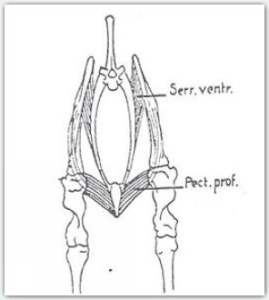
I demonstrated to Leanne the flexion of he thoracic vertebrae hat was related at a specific moment, with the lowering of the neck. My right hand that was placed under the vertebrae at the cervico-thoracic junction, provided a study support, mimicking the role of the serratus thoracis muscles. With the left hand. I lowered the cervical vertebrae. Leanne manipulated by herself the vertebral column feeling very clearly the neck position that was creating longitudinal flexion of the thoracic vertebrae.
I directed then Leanne attention on the fact that if she was giving a little more support with her right hand under the cervico-thoracic junction, exactly like if the serratus ventralis thoracis and cervocis muscles were giving greater support, the instant where the lowering of the neck was inducing flexion of the thoracic spine, will occur earlier. Leanne manipulated the vertebral column several times feeling the moment where the neck posture was having an effect on the thoracic vertebrae. Leanne also observed that without the support of her right hand under the cervico-thoracic junction, she did not felt the flexion of the thoracic spine related with the lowering of the neck. She experimented several time with and without support feeling very clearly that without the support of her right habd and therefore, without adequate suppodt of the trunk from the serratus evntralis thoracis muscles, the lowering of the beck only created a sagging of the whole structure without dynamic advantage in the thoracic vertebrae.
As we furthered our conversation on the matter, I told Leanne, “If you remember Chazot picture where he is walking on lose reins outside of the property, the neck position that he spontaneously selected is the neck position where this dynamic 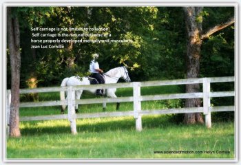 phenomenon occurs for him. As you can see, it is long but not low. He does have strong extrinsic forelegs muscles and in particular the serratus ventralis thoracic and he is therefore comfortable sustaining his trunk relatively high between his shoulder blades. If he had weaker serratus muscles, he would probably find comfort into a slightly lower neck posture. However, if I was asking for a lower neck posture, I would place him out of his comfort zone, his neck would no longer enhance his vertebral column mechanism and I would increase the weight on his forelegs.
This silhouette is the exact copy of a picture that was presented promoting lowering of the neck. The horse is at the trot. His left diagonal, right hind leg and left foreleg should be both into the swing phase. Instead look at the right hind limb. The hoof is clearly off the ground, which is normal at this sequence of the stride. By contrast, look at the left front leg. 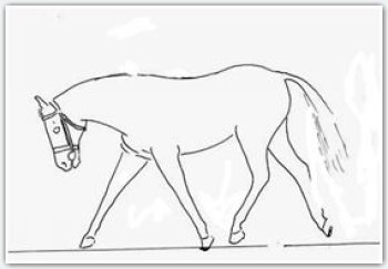 The limb is still heavily loaded on the ground. The hoof should be as much if not more off the ground that the right hind. The right front limb is going to impact before the left hind leg and in fact probably before the push off of the left front leg.
On the original picture there is a rider and you can easily imagine the discrepancy between what the rider believes that she is doing, “stretching, swinging, relaxed,” and what really happened. The horse is dysfunctional exhibiting aberrant kinematics. Lameness is around the corner.
Of course, when the neck is not forced into a low neck posture, upper neck muscles and the nuchal ligament are enhancing the dynamic action of the head and neck on the thoracic vertebrae. I asked Leanne if she remembered the video that we have published in one of the functional anatomy documents of the IHTC. The document showed a manipulation made on the nuchal ligament in the necropsy room. Leanne remembered that the specimen was lying on a table and we were showing the effect of the nuchal and supraspinous ligament when the neck was placed on a longer posture. Without the support given by the table, the greater tension of the nuchal ligament would have simply increase the sagging of the trunk between the forelegs and the concavity of the cervical vertebrae lower curve.
A long neck posture with appropriated developments of the extrinsic forelegs muscles and in particular the serratus system does have positive effects. Placing the neck lower and making up theories as inaccurate as “stretching” and as stretched as “mental peace,” might satisfy the rider’s mind but alters the horse talent and damage its physique.
Jean Luc
. Jean Luc Cornille 2013


