Mechanoresponsiveness PT 5
Mechanoresponsiveness
Part 5
Sclerotic thickening of the subchondral bone is a major factor in the pathogenesis of arthritis. Excessive loading and the accumulation of microdamage are thought to be important elements in the development of arthritis. Unless one possesses a Phillips XL30 scanning electron microscope, no one ever see microdamages or even microfractures and therefore accept candidly riding and training techniques increasing the load on the forelegs, lowering of the neck, driving the horse onto the bit, more forward, etc.
We often state, if a riding or a training technique is not good enough for the higher level of performances, it is not good enough for lower level horses. Same can be said about scientific knowledge. A little bit of science might be good enough to accredit simplistic theories, but if we look deeper, the magnitude of the damages questions the value of popular theories. Loading of the forelegs is the main reason for all forms of arthritis. “Subchondral clerosis and area of bone devitalization are common in the overload arthrosis of equine athletes.” (R.W. Norrdin and S. M. Stover, Subchongral bone failure in overload arthrosis: A scanning electron microscopic study in horses, 2006; 6(3); 251-257). ) Before listening and even believing in riding and training techniques loading the horse front legs one might need to see how microcracks evolve into subchondral sclerosis and area of bone devitalization. 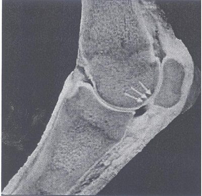
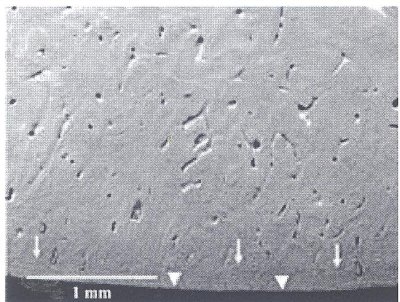
Thanks to the fascinating work of R. W. Norrdin and S. M. Stover and also the wonders of modern technology, here is how an unaffected subchondral bone looks like
Unaffected palmar subchondral bone site from a sample in the group of horses with mild sclerosis. Non-calcified articular cartilage is present at the bottom (arrowhead). The interface of the calcifies cartilage and bone can be seen in places (arrows)
The next picture shows subchondral bone with microfracture lines and early collapse
Subchondral 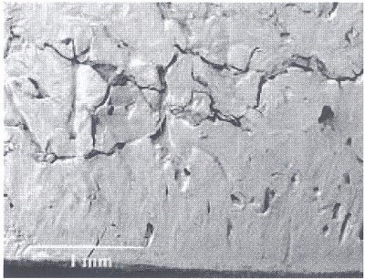 bone with microfracture lines and early collapse in a sample from the group with severe sclerosis and focal rarefaction. Note multiple cracks and fragmentation of bone with formation of gaps within fracture lines.
The next picture shows a higher magnification of the developing microfractures observed on the previous picture.
Higher magnification of arca along the developing microfracture in the previous picture. Note mismatched surfaces (arrows) and fragmentation of margins indicating fracture gap has collapsed. Osteoclastic erosion sites (arrowheads) were seen in larger developing cracks and indicated there had been remodeling at the site. 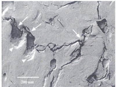
Bones do have a remarkable capacity of remodeling but if stress (excessive load on the forelegs) alters the bone remodeling capacity, the development of microcracks in the subchondral bone leading to arthritis in the cartilage is evolving right under your eye.
The next sample is from a group with advanced lesions showing gap in defect containing smoothly ground fragments of subchondral bone. The area in the white rectangle shows that there has been activated remodeling around the more chronic defect.
Incompletely ground sample from group with advanced lesions shows gap in defect containing smoothly ground fragments of subchondral bone. 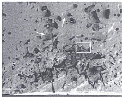 Enlarged vascular canals and resorption sites (arrows) indicate there has been activated remodeling around the more chronic defect.
The region of interest, which is the area within the rectangle is magnified in the next picture. The magnification shows an entrapped vessel. 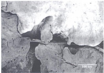
Higher magnification of microfracture gap with entrapped vessel. Note smooth edges on ground fragments indicating chronic wear on the surface.
The mechanism varies with the specific joint depending on the forces involved and the pattern of loading. This picture series have been made on racehorses. The lesions presented on these pictures is referred to as “Traumatic osteochodronsis” The problem is common on race horses. For many, the subchondral bone damages are asymptomatic.” However, the more sever forms are associated with fetlock lameness. Dressage and jumpers are subjected to less intense but similar overloading and when the riding and training technique reiterate overloading every training session, the bone remodeling does not repair the damages and the evolution that you are watching is leading the horse to all form of arthritis.
“Unload it” is the title of a recent study on human knee arthritis. Unloading it is how we restored soundness on Caesar, which suffered from arthritis between the second phalange and the coffin bone of the left hind leg. Unloading it is how we gave back her mobility and her life to the beautiful Diva, which suffered from double ring bones. Unload it is how we restore soundness on Manchester who suffered from both a stifle problem and arthritis on the same joint than Caesar but the other front leg. And the list goes on and on and on.
Since no one can see it until it is too late, it is easy to ignore the problem and keep lowering the neck and loading the horse front legs. The rider’s knowledge is the horse’s sole protection and therefore we continue the picture series further showing the evolution of the damages.
Subchondral bone microfracture at the site of collapse and indentation of cartilage (arrowheads) in a sample from the group with advanced lesions. 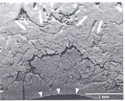 A fracture line through the calcified cartilage, (lower left) is associated with further infolding. The layer of bone above the microfracture has multiple fragmentation lines and a compacted appearance (outlined by arrows).
A closer view is underlining the reality of the damages.
Higher magnification of subchondral bone microfracture with collapsed matrix from the previous picture. Bone matrix above the microfracture line has multiple fine cracks (arrowhead) and a compacted arrangement. Also note continued fragmentation of fracture margins in the gap. 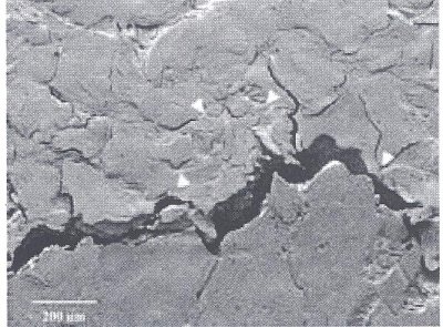
I
It might be interesting ending this article placing side by side a horse working in balance and a horse heavily loaded on the forelegs.
Horse in Balance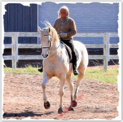
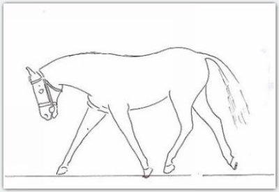
This is an outline of a photo of a horse
hevily loaded on the forehand
Jean Luc


 twitter
 facebook
 google
 stumbleupon
 pinterest
 linkedin